QIAGEN logo

Virtual Poster/PowerPoint Presentation Gallery

Presentation #1 (Poster)

teaser-6

Mechanistic insights into translational sciences

Stefanie Julia Willmann
Consultant and Research Participant, Helmholtz Zentrum München, TU München, Institute of Diabetes Research

Abstract: Translational sciences keep up the big picture beyond the deep-dive in diverse disease areas. Heterogeneity in dynamic cell populations and tissue morphogenesis in evolving organism, here represented by the evolving pancreas in the model of mus musculus, are ideal candidates for investigating precursor versus mature cells for the variants in the biologic dogma content, respective RNA versus DNA versus Protein. The gene Synaptotagmin 13 represents a candidate for translational sciences, as the gene itself suggests to be involved in pancreatic organogenesis and cancerogenesis. Preliminary results are pointing to phenotype in type 2 diabetes (T2D) in mus musculus along with a phenotype in the establishment of pancreatic duodenal adenocarcinoma (PDAC) in mus musculus.

See attached poster #1

Researcher email for contact: SJWillmann@gmx.de

Presentation #2 (PowerPoint)

T cell repertoire analyses in anti-PD-1 treated cancer patients

S. Simon1,2,3, V Voillet2, V Vignard1,3,4, Z Wu5, A. Khammari1,3,4, O Adotevi6, S. Rulli5, R. Gottargdo2, P Nghiem7, B. Dreno1,3,4, A. Riddell2, N. Labarrière1,3
1 University of Nantes, Inserm, CRCINA, F-44000 Nantes France
2 Fred Hutchinson Cancer Research Center, Seattle, WA USA
3 LabEx IGO "Immunotherapy, Graft, Oncology", Nantes, France
4 CHU of Nantes, Nantes, France
5 QIAGEN Sciences, Frederick, MD, USA
6 University Bourgogne Franche-Comte', INSERM, EFS BFC, UMR1098, France
7 Division of Dermatology, Department of Medicine, UW School of Medicine, Seattle, WA USA

Abstract: Clinical benefit from PD-1 inhibitors relies on reinvigoration of endogenous anti-tumor immunity. Nonetheless, robust immunological markers, based on circulating immune cell subsets associated with therapeutic efficacy are yet to be validated. We documented that the frequency of circulating PD-1+TIGIT+ (DPOS) CD8+ T-cells after 1 month of anti-PD-1 therapy was associated with clinical response. This DPOS T-cell population was enriched in highly activated T-cells, tumor-specific and emerging T-cell clonotypes and T lymphocytes overexpressing CXCR5, a key marker of the CD8 cytotoxic follicular T cell (Tfc) population. Additionally, transcriptomic profiling defined a specific gene-signature for this population as well as the over-expression of specific pathways associated with the therapeutic response. Our results provide a convincing rationale for monitoring this PD-1+TIGIT+ circulating population as an early cellular-based marker of therapeutic response to anti-PD-1 therapy.

Download PowerPoint

Researcher email for contact: nathalie.labarriere@inserm.fr

Presentation #3 (Poster)

miRNA Profile Assessment of Urine Exosomes from Boric Acid Treated Rats as Potential Biomarkers for Testicular Toxicity

R. Wayne Ball1, Barbara Gould2, and Jason Wise2
1 REACH Borates Consortium, Brussels, Belgium
2 QIAGEN Inc., Germantown, MD

Abstract: The objective of this study is to identify miRNAs in urine specific to boric acid (BA) related reproductive effects in rats. Urine samples were evaluated to identify differentially expressed testicular-specific miRNAs in BA treated rats using next-generation sequencing (NGS). Boric acid was administered to male Sprague Dawley rats via oral gavage at a dose level of 500 mg BA/kg bw/day for 28 days, a dose level known to produce fertility effects in male rats with minimal overt signs of toxicity. At the end of 28-days urine was collected over 24 hours. Urine samples were shipped to QIAGEN Genomic Services for exosomes isolation and miRNA analysis. Histopathology was conducted on testes and epididymis confirming BA treatment related effects. Test substancerelated findings included lower epididymis weights, smaller epididymides, and microscopic findings of cellular debris and decreased spermatid cellularity in the epididymis and tubular degeneration/atrophy and atypical residual bodies in the testis. Tubular degeneration/atrophy in the test substance-treated group was characterized by decreased numbers of germ cells with 
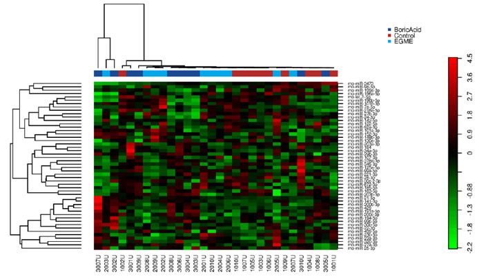
degeneration of spermatocytes and spermatids. Urine exosomes and miRNA were isolated with QIAseq 52 Spike-ins through exoRNeasy. miRNA sequencing was performed using an Illumina NextSeq500. The miRNA library preparation was completed using the QIAseq miRNA Library Kit. Several miRNAs were identified in rat urine as possible biomarkers for BA related effects on male reproductive system: miR-34c-5p, miR-449a-5p and miR-122-5p were decreased in BA treated rats compared to untreated controls. These miRNAs have also been identified to be decreased in humans with sertoli cell related spermatogenic failure. BA has been shown to affect Sertoli Cells in the testes of rats. Additionally, let-7-5p (decreased), mir-141-3p (increased) and miR21-5p (increased) were differentially expressed in BA treated rats. These miRNAs have been identified as potential biomarkers for human male non-obstructive azoospermia. Also, miR-27b-3p levels decreased in BA treated rats shown to be associated with asthenozoospermia in humans. These results provide the basis for the potential application of miRNAs as biomarkers for BA related testicular toxicity and highlight the value of urine exosome miRNA NGS as a discovery tool.

See attached poster #3

Researcher email for contact: barbara.gould@qiagen.com